
Coloured Samples in Chromogenic Endotoxin Testing: Why 405 nm Assays Fail and How αBET™ Helps

Colour is observed when some part of the visible spectrum is absorbed when white light is incident on a surface. Light that is not absorbed is either reflected or transmitted, minus the absorbed wavelengths, giving rise to colour1. For instance, when light in the range of 400–450 nm (blue/violet) is absorbed, the reflected or transmitted light appears yellow. Similarly, if a sample appears blue, it has absorbed light in the yellow/orange part of the visible spectra (570–600 nm).
All FDA-licensed chromogenic LAL assays use 4-nitroaniline (or para-nitroaniline [p-NA]) as the reporter chromophore. This is also true for the newer recombinant assays. All chromogenic reagents work identically by measuring the absorbance of the free form of p-NA as it is cleaved from a synthetic peptide (typically Boc-Leucine-Glycine-Arginine) by an enzyme that becomes active only in the presence of endotoxin.su
The key to the assay is that the absorbance maximum of unconjugated p-NA is around 381 nm, whilst the peptide-conjugated form has an absorbance maximum at ~315 nm. Typically, measurements are made at ~405 nm where the contribution from the conjugated form is virtually zero. Therefore, measuring the appearance of unconjugated p-NA at 405 nm is an effective way of monitoring the progression of the endotoxin response—the more rapid the appearance of p-NA, the greater the concentration of endotoxin in the sample.
If the sample is coloured, this can negatively impact the sensitivity of the assay2. For instance, if a sample appears yellow, it absorbs light in the UV/blue region of the visible spectra (350–450 nm), which overlaps the analytical wavelength for p-NA-based chromogenic assays (~405 nm). Therefore, as sample colour intensifies and background absorbance increases, the difference between background and reaction endpoint absorbance becomes increasingly small and eventually the assay endpoint becomes unresolvable from background absorbance, and the assay fails (Figure 1). Whilst this does not necessarily mean chromogenic reagents should not be used with coloured samples, it does mean, that along with endotoxin limit, endotoxin recovery, maximum viable dilution and low-endotoxin recovery, sample colour must be part of the decision-making process when selecting the most appropriate reagent and optimising an endotoxin assay.
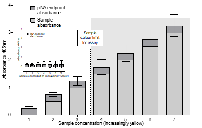
Figure 1. Endpoint absorbance is cumulative of sample and p-NA absorbances. When the sample concentration reaches ‘4’, the error in the endpoint measurement is greater than the magnitude of the maximum p-NA absorbance, and the assay fails. The magnitude of the error in the endpoint value is independent of whether the starting point of the assay is auto-zeroed or not.
Figure 1 illustrates the problems associated when working with yellow samples, but such issues are not limited to this colour space. Green samples are known sources of interference for p-NA-based assays. Furthermore, with the absorbance spectra of some materials tailing down to 400 nm, orange samples can also interfere with assays run at 405 nm. In addition, darkly coloured samples of any hue can impact p-NA assays given that even small background absorbance bands can become significant at very high analyte concentrations.
How does αBET™ overcome this problem?
αBET™ is a fully integrated and Pharmacopoeia compliant endotoxin testing platform delivering a greatly reduced time-to-result while demonstrating superior resistance to many of the optical interferences that plague market-leading rapid testing systems, tube readers, and plate readers3.
This unique level of robustness arises from the pairing of CMD proprietary 850 nm near-infrared (NIR) magneto-optical detection with FUJIFILM’s endotoxin-specific Pyrostar turbidimetric LAL reagent. By shifting optical interrogation into the NIR, common UV/visible-light optical interferences become irrelevant, allowing true endotoxin detection with minimal to no sample dilution. The αBET™ range includes a single port (4 samples) master instrument, a single port extension module and, in Q4 2026, for users requiring higher throughputs, a 4 port (16 sample) version of the master instrument will become available.
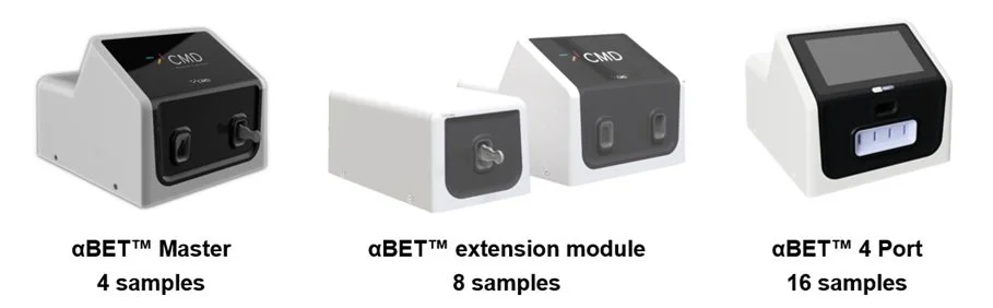
The huge advantage of working in the NIR is that coloured and light scattering samples that absorb or strongly scatter around 405 nm, becomes ‘near transparent’ when working at 850 nm. The implications of this for endotoxin testing is profound in that the need to heavily dilute coloured and complex samples to overcome optical interferences is much reduced or negated. This allows the analyst to use dilution as a tool to adjust sample endotoxin concentration so that it lies mid-way in working range of the assay where precision, accuracy and reliability are greatest.
Why was the myth of testing coloured samples not debunked’?
In a 2023 publication (American Pharmaceutical Review, "The Myth of Testing Coloured Samples: Debunked")4, the authors chose to demonstrate the robustness of a standard p-NA chromogenic assay to sample colour by testing a methionine, inositol, and choline (MIC) injection using a plate reader at 405 nm. Whilst a standard MIC injection is colourless, it is commonly co-formulated with Vitamin B12 (cobalamin), which imparts a strong orange/yellow colour, as was the case in the 2023 study (Figure 2).
The authors demonstrated that while the chromogenic LAL assay produced invalid results with undiluted and 1:10 diluted samples, valid results could be obtained with samples diluted 1:100 and beyond. At 1:1000 dilution, the solution was close to colourless.
The critical caveat regarding cobalamin is that while it appears yellow to the eye, it does not absorb strongly around 400 nm; its strongest absorbance is actually at ~360 nm, well below the detection wavelength used in the study. It is therefore unsurprising that valid results were obtained for MIC solutions at a 1:100 dilution. However, it should be noted that even with near "water-like" absorbance at 405 nm, it was still necessary to dilute the sample 100-fold to overcome baseline interference. Should another sample type equally as intensely coloured as the one in the publication absorb directly at ~400 nm, it is highly unlikely that a valid assay could be obtained at dilutions less than 1:1000. This proves that the human perception of colour can be highly misleading in optical assays. If the authors had chosen a different yellow substance for their studies, their conclusions would have been starkly different. For instance, in contrast to "yellow" MIC, both riboflavin and tartrazine produce strongly yellow solutions that absorb strongly around 400 nm.
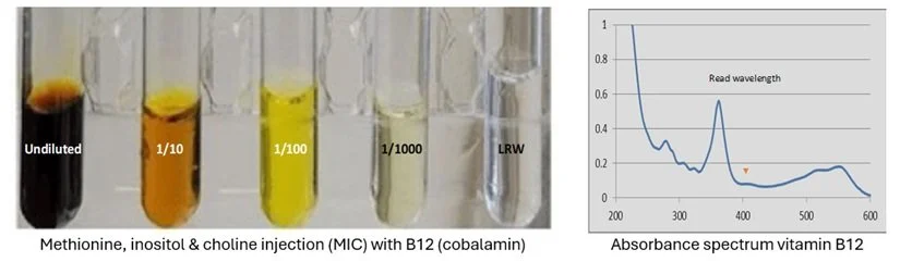
Figure 2. The myth of testing samples that are coloured debunked critique: Methionine, inositol, and choline injection (MIC) with vitamin B12 (cobalamin) and the corresponding absorbance spectrum of vitamin B12. (Source reference: https://www.a3p.org/en/myth-testing-colored-samples-debunked)
References
Kurt Nassau, (2001). The Physics and Chemistry of Color: The Fifteen Causes of Color. 2nd Edition. Wiley Interscience.
Samples that interfere with endotoxin assays: a modern dilemma. https://www.cm-dx.com/news-and-resources/samples-that-interfere-with-endotoxin-assays-a-modern-dilemma
Introducing the αBET™ system. https://www.cm-dx.com/alphabet
The myth of testing coloured samples debunked (2023). American Pharmaceutical Review. https://www.a3p.org/en/myth-testing-colored-samples-debunked